Weixin Service

 DouYin
 KuaiShou

Structural composition of electron microscope SEM
Date:2023-07-20 15:11:00 Views:2142
Electron Microscope (EM) is a type of microscope that utilizes electron beams for imaging. Compared to optical microscopes, electron microscopes have higher resolution and larger magnification. Electron microscope can be divided into Transmission electron microscopy (TEM) and Scanning electron microscope (SEM). This article will focus on the structural composition of SEM.
The main components of SEM include electron guns, focusing systems, scanning coils, sample stages, detectors, and imaging systems. Among them, the electron gun is the core component of SEM, responsible for generating electron beams. The electron gun usually uses a hot cathode electron gun or a field emission electron gun, and the generated electron beam passes through the focusing system and scanning coil, forming a high-density electron beam on the surface of the sample, thereby interacting with the sample surface. The function of the focusing system and scanning coil is to focus and scan the electron beam, enabling it to form a high-resolution electron beam on the surface of the sample.
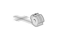
_20230720145102_663.jpg "电子显微镜sem的结构组成")
The sample stage is another important component of SEM, used to fix and adjust the position and angle of the sample. The sample stage of SEM usually has multiple functions, such as rotation, tilting, lifting, and rotation, which can enable samples to be imaged and analyzed at different angles. In addition, the sample stage can also process the sample through heating or cooling methods for more accurate analysis and observation.
The detector is a signal detection and imaging system of SEM, which is used to detect the Secondary electrons or backscattered electron signal generated on the sample surface and convert the signal into an electronic image. The detectors of SEM usually include Secondary electrons detector, backscattered electron detector and energy spectrum analyzer, which can conduct high-resolution imaging and chemical analysis on the sample surface.
The imaging system is another important component of SEM, used to convert the signals detected by the detector into electronic images. The imaging system of SEM usually includes signal amplifiers, scanning controllers, digital converters, and displays, which can perform high-resolution imaging and analysis of the sample surface.
In summary, the structural composition of SEM includes multiple parts such as an electron gun, focusing system, scanning coil, sample stage, detector, and imaging system. Each part cooperates with each other to achieve high-resolution imaging and analysis of the sample surface. The advantages of SEM such as high resolution, high sensitivity, and chemical analysis make it widely used in fields such as materials science, biology, geology, electronics, etc.